
I
Cornea
The clear front window that bends incoming light.
- Conditions: Infections, keratoconus, dry eye
- Tests: Topography, slit-lamp
Compassionate consultations, advanced diagnostics and specialist-led treatment for cataract, cornea, retina, glaucoma and complete family eye care.
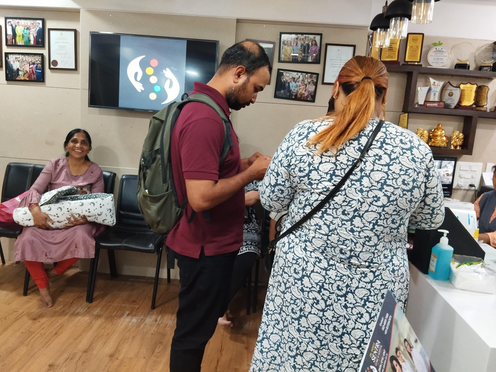
Welcome to clarity
At Sentra Clinic & Hospital, every consultation starts with your story — your symptoms, your work, your worries. Only then do we examine, measure and explain. Our Mumbai clinic pairs specialist expertise with advanced diagnostics, so decisions about your eyes are made with evidence, not haste.
Symptom navigator
Select a symptom to see general educational guidance and the right specialty to consult.
Guidance and the relevant specialty will appear here.
Disclaimer: This tool does not diagnose medical conditions. For sudden vision loss, severe pain, chemical injury or flashes with floaters, seek immediate medical evaluation.
Complete ecosystem
Hover or tap a specialty to explore it.
Our team
Specialist knowledge with a patient-first approach.

Photographs and credentials are published only after verification by the hospital.
Eye education
Follow the path of light through the eye — and learn what can go wrong at each stage.
The clear front window that bends incoming light.
The focusing lens that fine-tunes the image.
The light-sensing layer that creates the signal.
The cable carrying the signal to the brain.
Educational information only — not a diagnosis. Consult an eye specialist for personal advice.
Signature care pathways
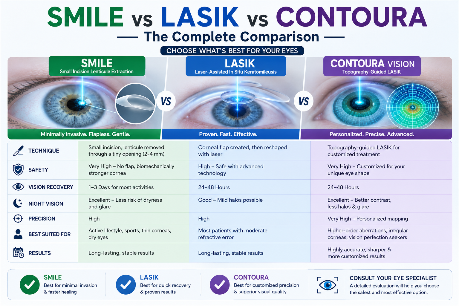 A beam of light,
Consultation, structured suitability assessment and honest treatment planning. We measure first — corneal shape, thickness, tear film and retina — then tell you clearly whether laser vision correction is right for your eyes.
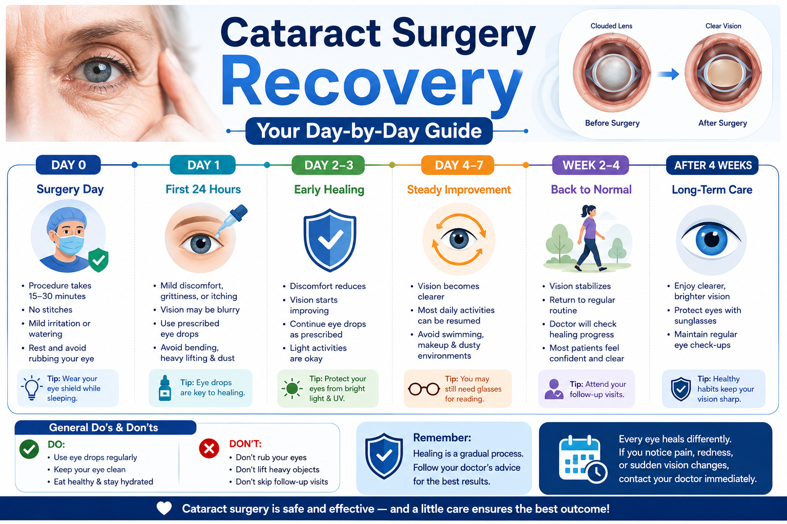 From cloudy
A cloudy lens scatters light and dims the world. We guide you through evaluation, intraocular lens choices and follow-up care — with realistic expectations, never guarantees.
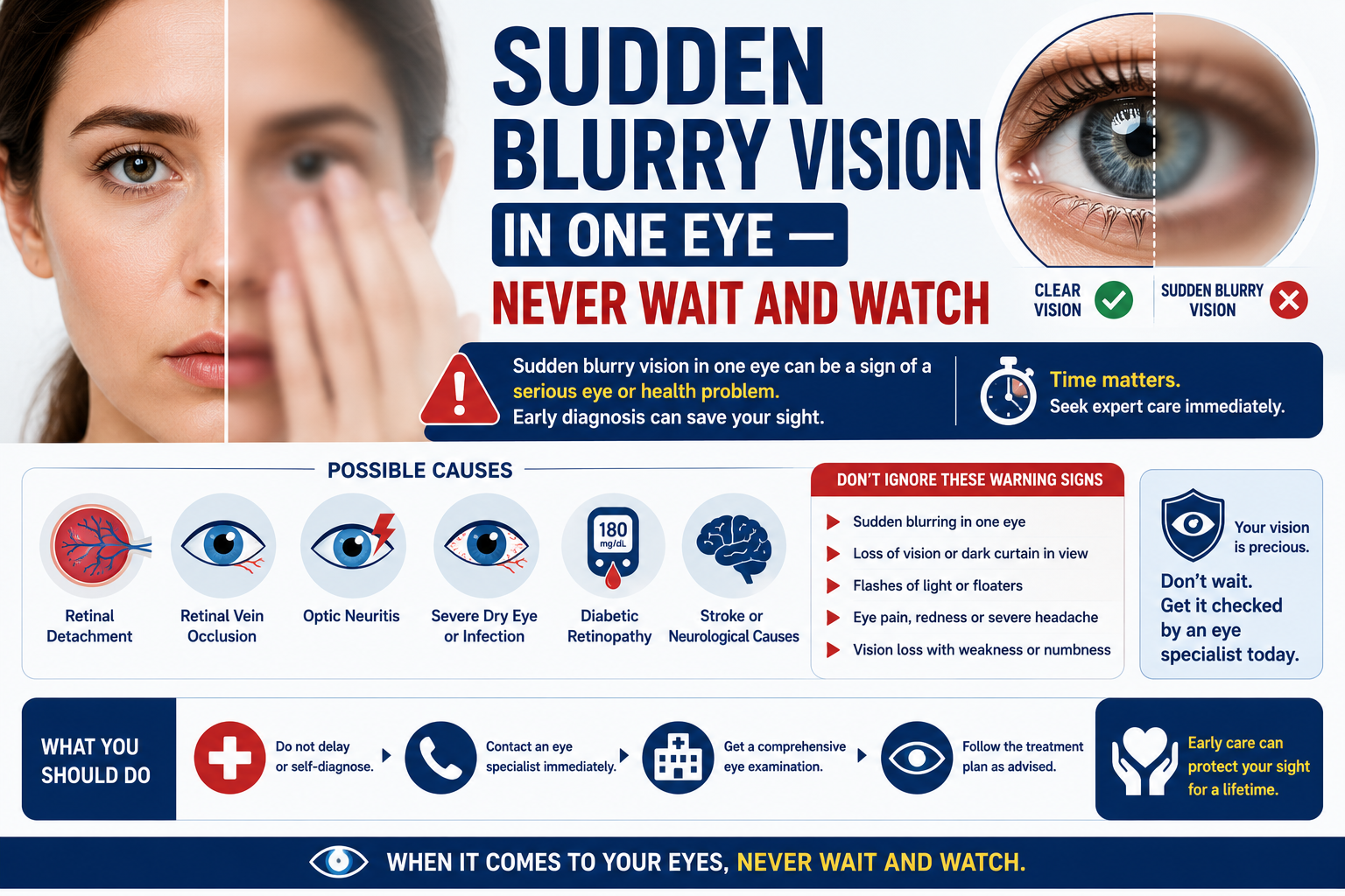 The retina,
Diabetic retinal screening, sudden flashes and floaters, macular conditions — the retina rewards timely assessment. Our imaging-led approach catches problems early and explains them clearly.
Diagnostic technology
A clean diagnostic pathway: what each test examines, why it may be recommended and what you can expect.
 Diagnostics
What it examines: Cross-sectional imaging of the retina and optic nerve layers.
Why recommended: Detects macular swelling, glaucoma-related nerve thinning and retinal disease before symptoms appear.
What to expect: Non-contact light scan; a few minutes per eye. Dilatation may be advised.
Diagnostics
What it examines: A curvature and elevation map of the cornea.
Why recommended: Essential for LASIK suitability, keratoconus detection and contact-lens fitting.
What to expect: Painless; you simply look at a target light for a few seconds.
 Diagnostics
What it examines: High-resolution photographs of the retina.
Why recommended: Documents diabetic changes, retinal tears and other findings for accurate comparison over time.
What to expect: A bright flash photograph; usually quick, sometimes after dilatation.
 Diagnostics
What it examines: Maps your field of vision point by point.
Why recommended: Detects and monitors glaucoma and neurological visual field loss.
What to expect: You press a button when you see lights; typically 10–15 minutes.
Diagnostics
What it examines: Microscope examination of the front and back of the eye.
Why recommended: The foundation of every eye examination — detects cataract, corneal and surface disease.
What to expect: You rest your chin on the microscope; completely non-invasive.
 Diagnostics
What it examines: Measures intraocular pressure.
Why recommended: A key screening and monitoring test for glaucoma.
What to expect: A gentle puff of air or a numbed touch measurement; seconds per eye.
From the clinic
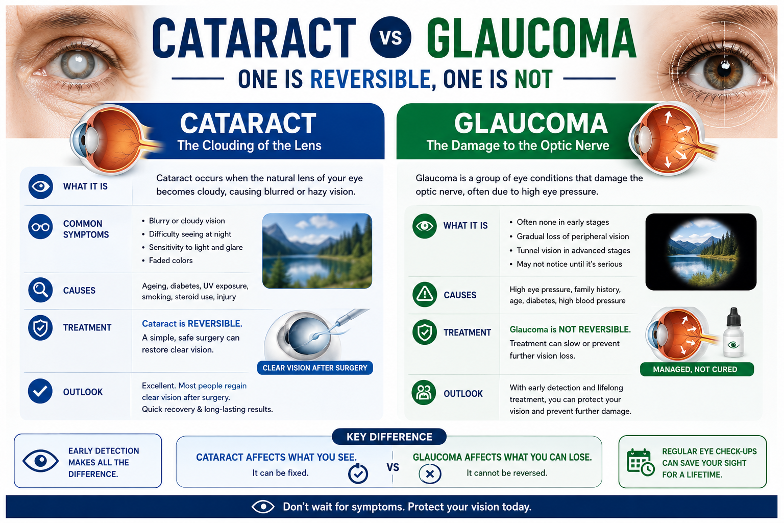
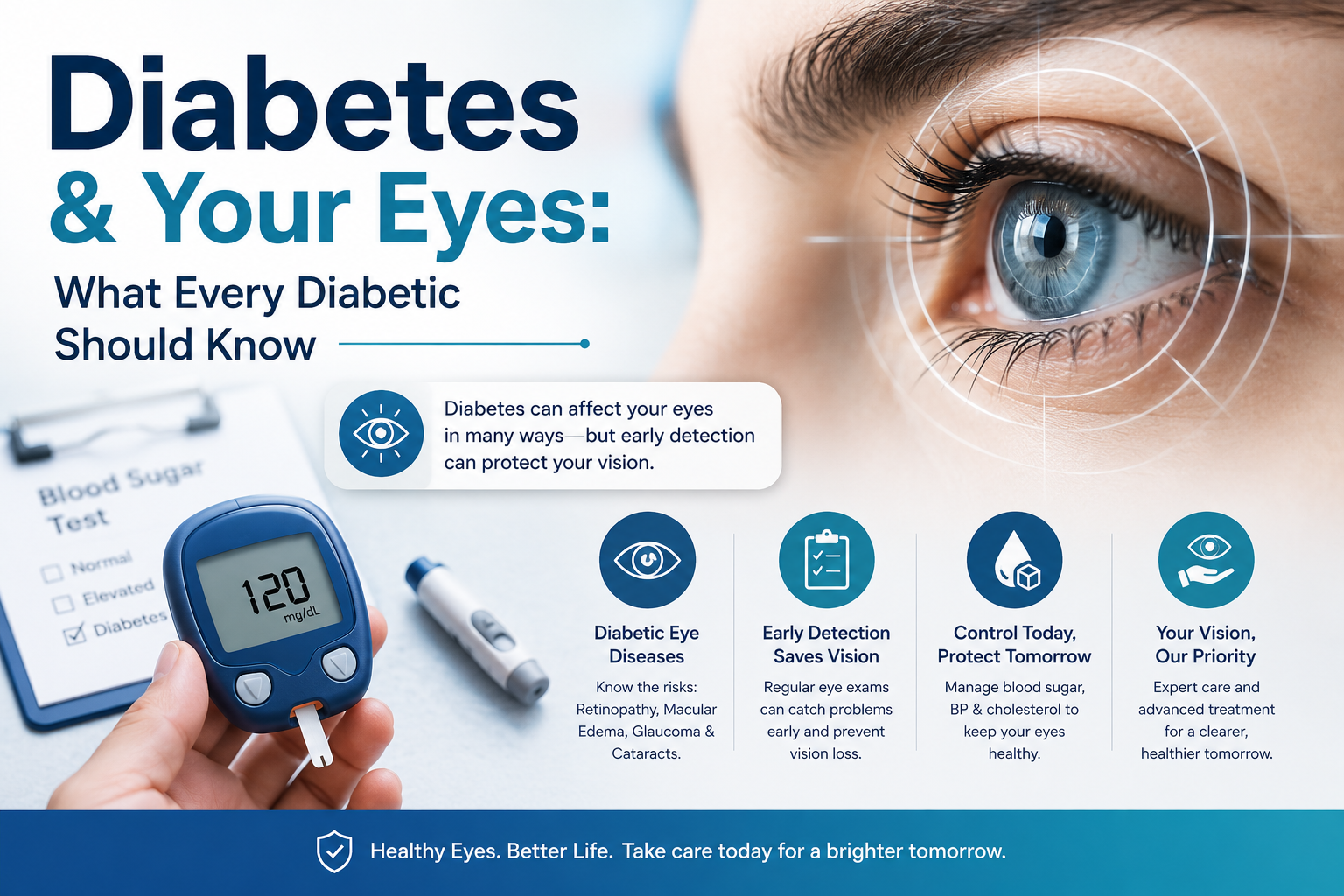
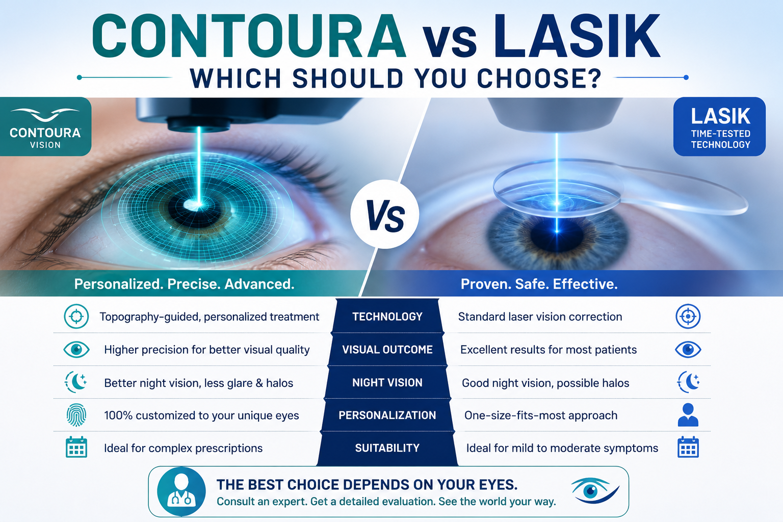
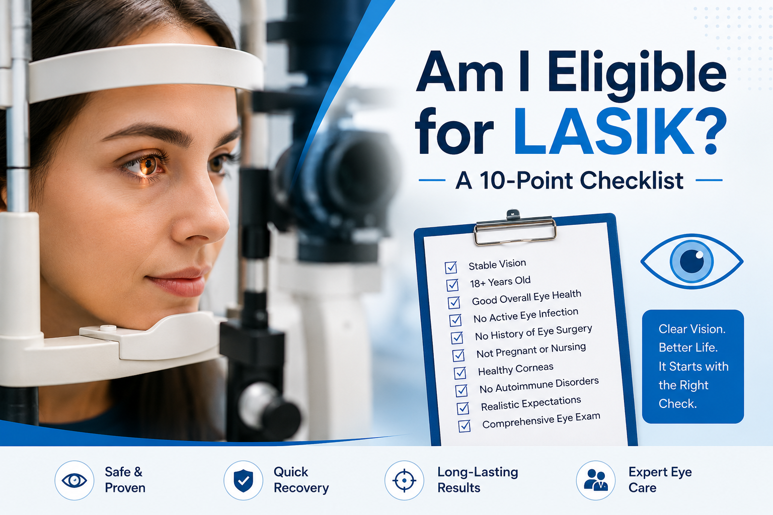
Explore the clinic-provided visual guides and care resources.
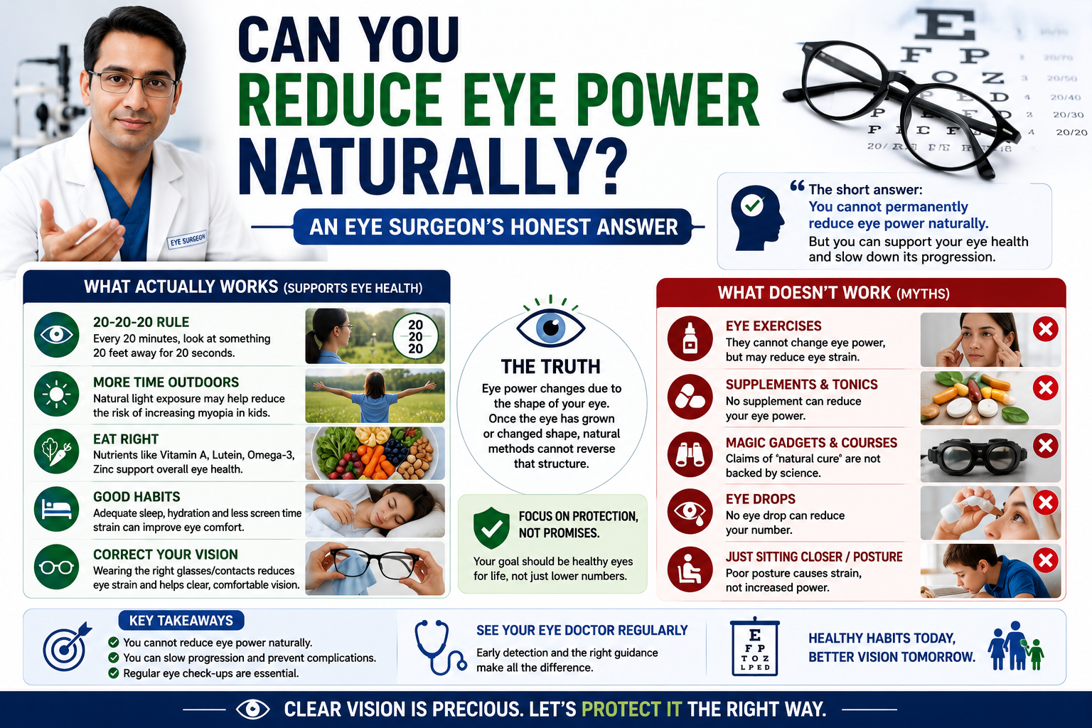
Practical education for clearer everyday vision.
Simple information to support regular eye care.
Know when retinal symptoms need prompt attention.
Understand why screening and follow-up matter.

Visual guidance for growing eyes and early checks.
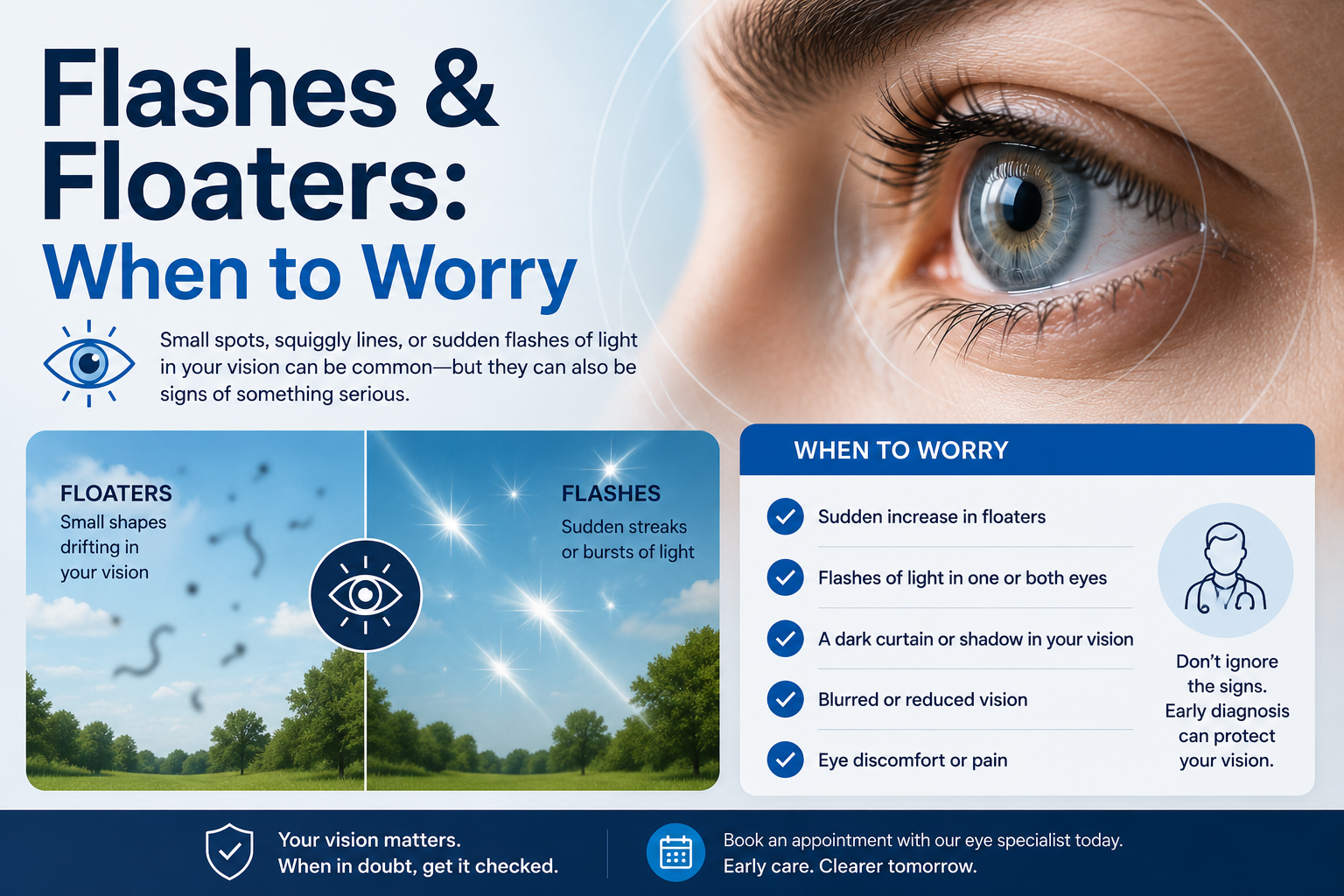
Learn about common symptoms and supportive care.
Helpful education for everyday eye decisions.
A visual guide to informed conversations with your specialist.

Clinic-provided information to review before a visit.
Keep the clinic-provided emergency contact information close.
First visit, made simple
Seven calm steps from enquiry to follow-up — so you always know what happens next.
Select a service
Book an appointment
Receive confirmation
Attend consultation
Complete diagnostic testing if advised
Discuss a personalized care plan
Receive follow-up guidance
Care standards
Structured sterilisation and clinical hygiene protocols across consultation and diagnostic areas.
Findings, options and costs explained before any decision — with time for your questions.
Scheduled reviews and clear post-visit guidance so recovery never feels uncertain.
Cashless or insurance assistance is shared once confirmed by the clinic. Please ask at reception.
Knowledge library
Most floaters are harmless — but a sudden shower of them, or new flashes of light, can signal a retinal tear. Here is how to tell the difference.
Read article →Cataracts develop gradually. Glare while night driving and frequent power changes are often the first clues.
Read article →Diabetic retinopathy often has no early symptoms. Annual screening catches damage while it is still highly treatable.
Read article →Appointments, first visits, children’s checks, emergencies — quick answers.
Browse FAQs →A downloadable “Preparing for Your Eye Visit” guide is being prepared.
Coming soonBook a visit